
 English
 日本語
日本語 한국어
한국어 中文(廣東話)
 Tagalog
 ไทย
ไทย Tiếng Việt
 Chuukese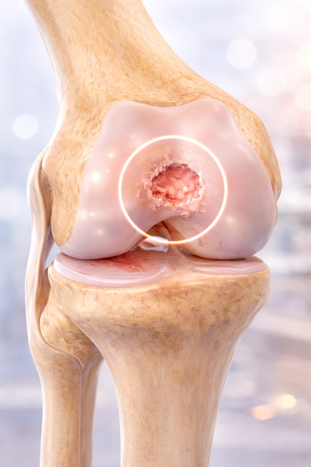
Cartilage restoration in Honolulu — the short answer
Cartilage restoration repairs a focal defect, a contained pothole in the knee's joint surface, so an otherwise healthy knee doesn't need a replacement. It is for active patients with a symptomatic, contained lesion in a knee still worth preserving, including selected knees with early arthritis. Dr. Morton offers the full ladder as outpatient surgery in Honolulu, including Hawai‘i's first arthroscopic MACI and CartiHeal Agili-C. Recovery takes months; individual results vary.
- Focal defect, not diffuse arthritis — the whole evaluation turns on that distinction
- Six rungs on one ladder, matched to defect size, depth, location, and your goals
- Agili-C is FDA-labeled for KL grade 0–3 arthritis and 1–7 cm² of treatable area — a "grade 3" X-ray alone no longer closes the door
- Evidence-backed: MACI beat microfracture in the randomized SUMMIT trial; Agili-C beat debridement/microfracture in a 251-patient randomized trial
- Outpatient surgery with PB&J Physical Therapy in Honolulu and Kunia; clinics in Honolulu, Kunia, Hilo, and Kona
What articular cartilage is — and why it can't heal itself
Articular cartilage is the smooth, glass-like surface that caps the ends of the bones inside your knee. It has one job, done superbly: reduce friction and spread load, so thousands of steps, squats, and waves paddled into leave the joint unbothered. But that performance comes at a price — cartilage has no blood supply of its own, and full-thickness cartilage damage has very limited ability to restore itself to normal, native hyaline cartilage. A deep defect doesn't scab over and heal the way skin or bone does. It persists.
That's why some cartilage injuries stay painful despite months of rest and rehabilitation. The typical signature: swelling after activity, deep aching in the joint, catching or clicking, trouble with stairs and squatting, recurrent effusions, and a knee that won't let you get back to hiking, running, or sport. But here is the honest counterpoint — cartilage abnormalities also show up on MRIs of knees that feel fine. Not every defect needs treatment, which is why Dr. Morton treats the patient plus the knee, never the MRI report alone.
The pothole and the road: restoration versus replacement
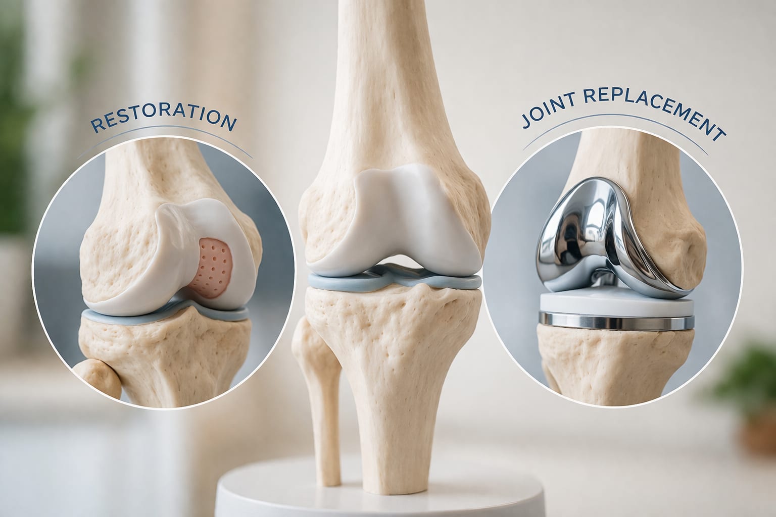
Think of the cartilage surface as a road. A focal defect is a pothole — a discrete, contained area of damage surrounded by healthy pavement. Diffuse osteoarthritis is a road worn out along its whole length. Cartilage restoration fills the pothole; knee replacement repaves the road. Choosing between them starts with a single question: is there a treatable cartilage or osteochondral lesion in a knee that is still worth preserving?
Older thinking sorted patients into two tidy boxes — focal defect or arthritis. Real knees are more complicated. A patient can have a clinically important focal lesion and radiographic osteoarthritis at the same time. Radiologists describe arthritis severity with the Kellgren-Lawrence (KL) scale: KL3 means definite bone spurs with definite joint-space narrowing; KL4 is severe, end-stage disease. For years, any mention of "grade 3 arthritis" ended the cartilage-restoration conversation. It shouldn't — at least not automatically. The FDA-approved indication for the CartiHeal Agili-C implant expressly includes knees with KL grade 0 through 3 osteoarthritis, provided the lesion and the rest of the knee fit the labeling. KL4 remains a contraindication, and diffuse advanced arthritis still favors arthritis-focused treatment — but a KL3 report alone no longer closes the door. It means the knee deserves a more detailed structural and mechanical assessment, not a reflexive verdict in either direction.
Who is a candidate? The evaluation looks far beyond the MRI
A cartilage-preservation evaluation is a structural and mechanical audit of the whole knee. Weight-bearing X-rays show joint-space loss, bone spurs, and how widely the arthritis is distributed. Full-length standing alignment films reveal whether bow-legged or knock-kneed alignment is overloading the damaged compartment. MRI characterizes the cartilage itself, the bone beneath it, the menisci, and the ligaments. And the physical exam answers the question imaging can't: does what the pictures show actually match this patient's pain and function?
From all of that, Dr. Morton works through the decision questions that determine whether restoration makes sense:
- Is the lesion focal enough to treat — and where exactly is it?
- Is the defect contained, with healthy cartilage shoulders around it?
- Is the bone beneath the defect healthy enough to build on?
- How much osteoarthritis exists elsewhere in the knee?
- Is the meniscus functional, and is the knee ligamentously stable?
- Is malalignment overloading the damaged compartment?
- Has appropriate nonsurgical treatment genuinely been tried?
- Do your activity goals justify the recovery and the risks?
First rung: no surgery at all — therapy, and where biologics fit
Not every cartilage defect needs an operation, and the ladder starts on the ground. Physical therapy can transform a painful knee by restoring motion, rebuilding quadriceps and hip strength, and retraining the knee's ability to tolerate load. Activity modification, weight management when appropriate, bracing in selected alignment patterns, and targeted injections round out the nonsurgical program. A successful course of rehab doesn't mean the cartilage regrew — it means the knee became functional enough that surgery was unnecessary. That is a win, not a consolation prize.
Orthobiologics sit on the same rung, honestly framed. Platelet-rich plasma (PRP) and bone marrow aspirate concentrate (BMAC) can calm inflammation and improve symptoms in early cartilage disease, and they can support the healing environment around a surgical repair — but no injection regrows a full-thickness cartilage defect, and Dr. Morton will tell you so plainly. His orthobiologics program explains what the evidence supports, and what it doesn't, before you spend anything on it.
When persistent swelling, catching, or activity-limiting pain continues despite a genuine rehabilitation effort, that is the signal to look harder at whether the cartilage lesion is truly driving the symptoms — and whether one of the surgical rungs can fix it.
The surgical ladder: debridement and microfracture
The simplest surgical rung is arthroscopic debridement, or chondroplasty — smoothing frayed, unstable cartilage flaps through keyhole incisions during knee arthroscopy so they stop catching and inflaming the joint. It doesn't grow new cartilage, but for the right mechanical symptoms it provides real relief with a short recovery.
One step up is microfracture, a marrow-stimulation technique: small perforations are made in the bone at the base of the defect, releasing marrow elements that form a repair tissue over the lesion. It is a time-tested, single-stage option for smaller defects — and, along with debridement, it served as the surgical standard-of-care comparator in the pivotal trial that led to the Agili-C approval. Its honest limitation: the repair tissue is fibrocartilage, a scar-like patch that is less durable than the native hyaline cartilage it substitutes for. For larger defects, or in patients with higher demands and more years to cover, the restorative rungs above it usually serve better.
OATS and osteochondral allograft: transplanting cartilage with its bone
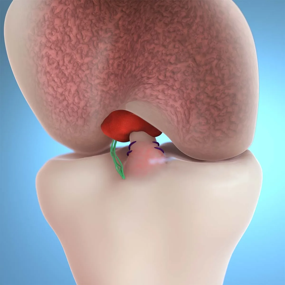
Some defects go deeper than the cartilage — into the bone that supports it. These osteochondral lesions are treated by transplanting both layers together.
OATS — osteochondral autograft transfer — moves small cylinders of your own cartilage and underlying bone from a lower-demand area of your knee into the defect. Its great advantage is biology: the defect is resurfaced with your own living hyaline cartilage, already attached to its own bone, in a single operation. Its honest limit is supply — only so much donor tissue can safely be harvested from one knee, which caps the defect size OATS can cover.
When the defect is larger, when substantial bone is involved, or when a previous cartilage procedure has failed, osteochondral allograft extends the same principle using precisely matched donor cartilage and bone. Size stops being the constraint, and the defect is rebuilt with mature, structural tissue — a workhorse option for the big reconstructions that smaller techniques can't reach.
MACI: regrowing cartilage from your own cells
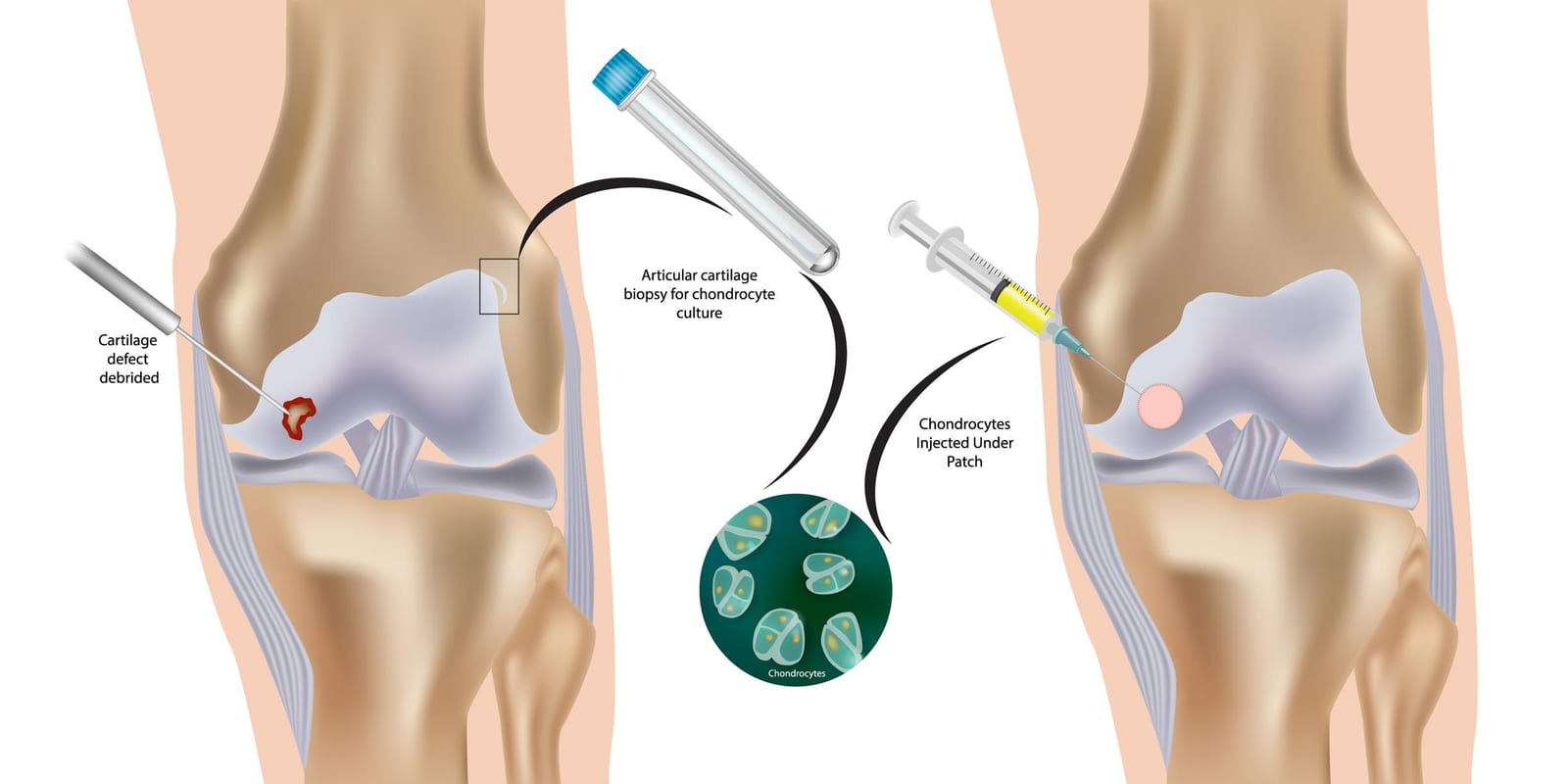
MACI — matrix-induced autologous chondrocyte implantation — is cartilage restoration built from your own cells. In a first, brief arthroscopic procedure, a small biopsy of healthy cartilage is taken from a non-weight-bearing part of your knee. Those cells — chondrocytes — are then grown and multiplied in a specialized laboratory and seeded onto a collagen membrane. At a second operation, the membrane is trimmed to the exact shape of the defect and implanted, where the cells mature into new cartilage tissue.
One more distinction belongs here: Dr. Morton is the first surgeon in Hawai‘i to perform arthroscopic MACI — implanting the cell-seeded membrane arthroscopically, through keyhole incisions, rather than through the larger open incision traditionally used for the implantation stage — keeping both stages of the treatment minimally invasive.
What the MACI evidence shows
Because the cells must be cultured, MACI is inherently a staged treatment — two procedures, separated by the laboratory phase. In exchange, it can resurface appropriately selected symptomatic full-thickness defects, including shapes and locations that plug-based grafts fit poorly — provided the joint around the defect remains preservable. It is a commitment: two operations and a disciplined rehabilitation arc. For the right defect in the right patient, it is also one of the most complete answers cartilage surgery has — and one of the best studied. In the randomized SUMMIT trial (Saris et al., American Journal of Sports Medicine 2014), 144 patients with symptomatic defects of 3 cm² or larger were treated with MACI or microfracture: MACI produced significantly greater improvement in pain and function at two years, with treatment failure in 12.5% versus 31.9%, and the advantage was maintained at five years (Brittberg et al., 2018). MACI has been FDA-approved in the United States since December 2016. Individual results vary.
CartiHeal Agili-C: the implant that widened the window
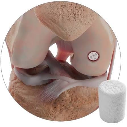
CartiHeal Agili-C is the newest rung on the ladder — an FDA-approved, cell-free, off-the-shelf, biodegradable scaffold made from aragonite, a natural form of calcium carbonate. It is biphasic: one zone engineered for the cartilage layer, one for the bone beneath it. Implanted into a prepared defect in a single operation, the scaffold is gradually resorbed as the body remodels it. It is not MACI — no cells are harvested or cultured — and it is not a knee replacement.
What makes Agili-C notable is its FDA-labeled indication: ICRS grade III or greater cartilage or osteochondral lesions of the knee, with a total treatable area of 1–7 cm², in patients with Kellgren-Lawrence grade 0–3 osteoarthritis. That last clause is the headline — the label expressly includes selected knees that already show mild-to-moderate arthritis on X-ray, rather than restricting the option to pristine joints. In the pivotal randomized study of 251 patients at 26 centers (Altschuler et al., American Journal of Sports Medicine 2023), Agili-C was compared against the surgical standard of care — debridement or microfracture. The implant group had superior scores at every follow-up: 77.8% were responders (at least a 30-point improvement in overall KOOS) versus 33.6% of controls, 88.5% had at least 75% defect fill on MRI at two years versus 30.9%, and the failure rate was 7.2% versus 21.4%. The FDA approved the device in 2022 on that evidence. Individual results vary.
Agili-C fine print: who it is not for
The fine print matters, and Dr. Morton reviews it candidly. KL4 — severe, end-stage arthritis — is a contraindication. The labeling also identifies situations where safety and effectiveness have not been established, several of them common in arthritic knees: significant malalignment, ligament instability, and absence of a functional meniscus. Lesion-specific limits apply too — uncontained defects, subchondral bone cysts deeper than 8 mm, and lesions within avascular necrosis — and the patella is not an indicated treatment site. Two patients can both carry a "KL3" label and receive completely different recommendations. That is not inconsistency; that is the evaluation working.
Cartilage doesn't fail in isolation: alignment, meniscus, and stability
A cartilage repair placed into a hostile mechanical environment is a patch on a pothole that the traffic keeps hammering. A bow-legged knee overloads the medial compartment. A deficient meniscus concentrates contact stress on the surface below it. An unstable knee — after an ACL tear, for instance — grinds abnormal shear across the joint with every giving-way episode, and kneecap instability does the same to the patellofemoral compartment.
Successful joint preservation therefore sometimes means treating the cause of the overload, not just the damaged surface: realigning the limb with an osteotomy, preserving or reconstructing the meniscus, or stabilizing the ligaments — occasionally in the same operation as the cartilage work. This is precisely why the evaluation examines the whole knee, and it matters most when restoration is being considered in a knee that already shows arthritis. Fix the pothole and ignore the traffic pattern, and the pothole comes back.
Recovery: measured in months, built to last decades
Cartilage restoration is outpatient surgery with an inpatient-sized commitment to rehabilitation — because the repair tissue needs time and protection to mature. The arc depends on the rung. A simple debridement recovers over weeks. Restorative procedures — microfracture, OATS, allograft, MACI, Agili-C — typically begin with a period of protected weight-bearing on crutches while the repair consolidates, paired with early motion exercises that nourish the healing surface. From there, rehabilitation progresses in stages: restoring full motion, rebuilding quadriceps and hip strength, then gradually reintroducing load. Return to impact activity and sport is measured in months, not weeks, and is gated by the tissue's biology and your progress rather than the calendar.
Dr. Morton's patients rehabilitate with PB&J Physical Therapy — the therapy practice he founded — at his Honolulu and Kunia locations, working from protocols matched to the specific procedure; neighbor island patients' rehab is coordinated with therapists near home, with telehealth check-ins along the way. The months of discipline buy something no artificial joint offers: a knee resurfaced with living tissue, with every future option still on the table.
- Surgery day
Outpatient procedure
Arthroscopic or small-open, home the same day with the knee protected.
- Early weeks
Protect and move
Protected weight-bearing on crutches for restorative procedures, paired with early motion that nourishes the healing surface.
- Then
Motion and strength
Full motion restored, quadriceps and hip strength rebuilt with PB&J Physical Therapy or a therapist near home.
- Months
Gradual loading
Load reintroduced in stages as the repair tissue matures.
- Months to a year
Impact and sport
Gated by the tissue's biology and your testing — not the calendar.
Recovery rung by rung
Rung by rung, the shape of recovery looks like this — timelines are individualized to your procedure and progress:
| Procedure | Operations | Early phase | Return to impact and sport |
|---|---|---|---|
| Arthroscopic debridement | One, arthroscopic | Walk the same day; crutches briefly for comfort | Weeks |
| Microfracture | One, arthroscopic | Protected weight-bearing on crutches with early motion while repair tissue forms | Months, gated by healing |
| OATS / osteochondral allograft | One | Protected weight-bearing while the bone plug incorporates, then progressive loading | Months, gated by imaging and strength |
| MACI | Two — biopsy, then implantation after laboratory culture | Protected weight-bearing and early motion after implantation while the cells mature | Months to a year, gated by tissue maturation and testing |
| Agili-C | One | Protected weight-bearing with early motion while the scaffold remodels | Months, gated by healing and testing |
When restoration is no longer the answer
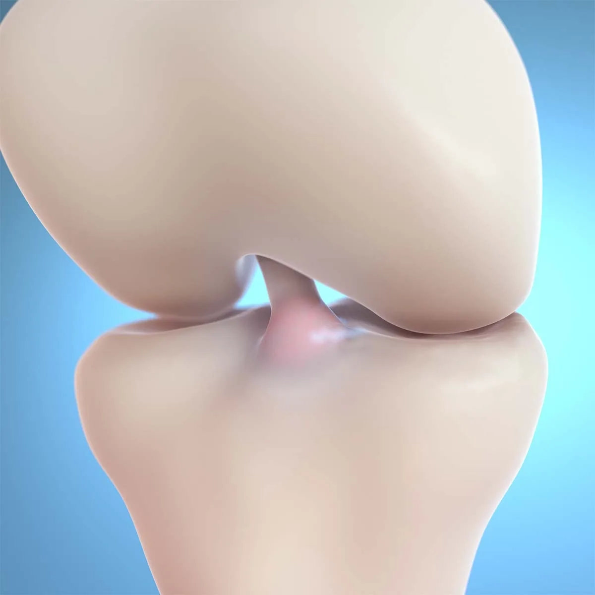
Cartilage restoration exists to preserve a knee — not to postpone replacement at any cost. When pain comes from diffuse, end-stage arthritis rather than a treatable focal lesion, patching one surface cannot address a whole-joint disease, and partial or total knee replacement becomes the more predictable strategy. KL4 arthritis is specifically contraindicated for Agili-C, and even before KL4, the overall pattern of disease can make arthroplasty the wiser choice for an individual knee.
This is where Dr. Morton's training changes the conversation. As a fellowship-trained adult hip and knee reconstruction surgeon, he performs the entire spectrum — from arthroscopic cartilage work to robotic partial and total knee replacement — so his recommendation is never shaped by what he happens to offer. A KL3 X-ray should not trigger either extreme conclusion: it doesn't automatically mean "too arthritic to restore," and it doesn't automatically mean "Agili-C candidate." It means your knee has earned a careful, honest assessment — and you will get exactly that, including a straight answer when the alternatives to replacement have genuinely run out.
The risks and limitations, stated plainly
Every cartilage procedure involves tradeoffs, and informed patients make better decisions. Depending on the operation, risks can include:
- Infection and blood clots — the baseline risks of any knee surgery
- Stiffness — one reason early, protocol-driven motion matters
- Persistent pain or swelling — not every repair silences every symptom
- Incomplete integration or graft/implant failure — repair tissue can fail to mature or bond
- Progression of osteoarthritis — restoration treats a lesion; it does not cure the disease process around it
- Additional surgery — a failed repair may need revision to another rung of the ladder, or to replacement
Agili-C carries its own labeled warnings, precautions, and potential adverse events, which Dr. Morton reviews with candidates as part of informed decision-making. And one limitation deserves italics: no cartilage-restoration procedure can guarantee that a knee will never need replacement. The honest goal is to make that day unnecessary — or at least distant — while keeping you active on your own joint in the meantime.
Where it happens — and how to start
Dr. Morton performs cartilage restoration as outpatient surgery at The Queen's Medical Center, at Adventist Health Castle, and at Hilo Community Surgery Center on the Big Island — the venue matched to your procedure, your health, and your insurance. Care is anchored by Pacific Bone & Joint, the practice he founded, with clinics in Honolulu, West Oʻahu (Kunia), Hilo, and Kona.
The starting point is an orthopedic consultation with weight-bearing X-rays — and your existing MRI, if you have one. It is an especially worthwhile visit if you've been told you are "too arthritic" for cartilage restoration based only on the phrase grade 3 osteoarthritis, or if you simply want a second opinion before committing to a replacement. Neighbor island and fly-in patients can begin with a telehealth visit and local imaging. Call (808) 439-6201 or book online. Bring a photo ID, your insurance card, and any prior X-rays, MRI, or operative reports; managed-care and Medicaid/Quest plans may require a referral from your primary care physician, which the office checks before your visit. You will leave with a diagnosis, honest options, and a plan that fits the actual pattern of your knee disease, whether that plan is rehabilitation, restoration, mechanical correction, or arthroplasty.
References
- Saris D, et al. Matrix-applied characterized autologous cultured chondrocytes versus microfracture: two-year follow-up of a prospective randomized trial (SUMMIT). Am J Sports Med. 2014;42(6):1384–1394. Source
- Brittberg M, et al. Matrix-applied characterized autologous cultured chondrocytes versus microfracture: five-year follow-up of a prospective randomized trial. Am J Sports Med. 2018;46(6):1343–1351. Source
- Altschuler N, et al. Aragonite-based scaffold versus microfracture and debridement for the treatment of knee chondral and osteochondral lesions: results of a multicenter randomized controlled trial. Am J Sports Med. 2023;51(4):957–967. Source
Medically reviewed by Paul N. Morton, MD, FAAOS, FAAHKS — board-certified orthopedic surgeon, fellowship-trained in adult hip & knee reconstruction and orthopedic trauma. Last reviewed: .
This page is for education only and isn’t a substitute for an exam and personalized advice — schedule a consultation to discuss your situation.