
The short answer: treat the damaged surface and the load pattern
Cartilage restoration addresses a focal area of damaged joint surface. Osteotomy addresses how force travels through the knee. When malalignment concentrates load in the same compartment as the defect, treating both may be considered so the repaired surface is not asked to absorb the same excessive stress.
That does not mean every bow-legged or knock-kneed knee needs bone realignment. Some alignment is constitutional and well tolerated. Osteotomy becomes a discussion only when the deformity, symptoms, cartilage pattern, meniscus, ligaments, age, activity goals, and arthritis distribution fit a joint-preserving strategy.
Why alignment changes cartilage loading
In a neutrally aligned leg, body weight is shared across the knee in a predictable pattern. A varus, or bow-legged, alignment commonly shifts more load toward the medial compartment. A valgus, or knock-kneed, alignment commonly increases load on the lateral compartment. A patellofemoral defect may have a different mechanical driver, such as abnormal kneecap tracking or an elevated tibial tubercle.
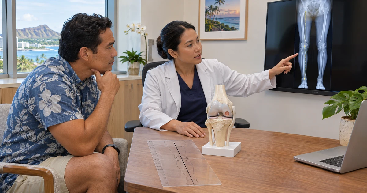
The location of the deformity matters. The visible angle at the knee does not reveal whether the source is mainly in the tibia, the femur, both bones, or the joint itself. Correcting the wrong level can create an oblique joint line or another mechanical problem. Planning therefore uses the entire standing limb rather than a short knee X-ray alone.
The evaluation uses more than an MRI
MRI describes the cartilage defect, the bone beneath it, the menisci, ligaments, and other soft tissues. It cannot replace standing mechanical assessment. A joint-preservation workup commonly combines symptoms and examination with weight-bearing knee X-rays, a full-length standing alignment film, and MRI when cartilage restoration is being considered.
- Does the pain localize to the same compartment as the cartilage defect?
- Is the lesion focal and contained, or is arthritis diffuse across the joint?
- Where does the weight-bearing axis pass through the knee?
- Does the deformity arise from the tibia, femur, both bones, or the joint?
- Is the meniscus present and functional?
- Are the ACL, PCL, and collateral ligaments stable?
- Is the knee able to fully straighten, and how much motion is present?
- Can the patient complete months of protected rehabilitation and bone healing?
High tibial osteotomy and distal femoral osteotomy are different tools
A high tibial osteotomy (HTO) changes the alignment of the upper shinbone. It is commonly used to unload an overloaded medial compartment in a varus knee, although the exact correction depends on where the deformity originates. A distal femoral osteotomy (DFO) changes the lower thighbone and is commonly used for valgus alignment with lateral-compartment overload.
Some deformities involve both bones and may require a more complex plan. Opening-wedge and closing-wedge techniques are available, and fixation generally uses a plate and screws while the bone heals. The operation is individualized; “straightening the leg” is not a one-size-fits-all target.
Which cartilage procedures may be combined with osteotomy?
Published series include osteotomy combined with marrow stimulation, osteochondral autograft transfer, osteochondral allograft, autologous chondrocyte implantation such as MACI, and other restoration approaches. The appropriate surface treatment depends on defect size, depth, location, containment, prior surgery, and the condition of the underlying bone.
Sometimes osteotomy is performed at the same operation as cartilage restoration. Sometimes it is staged. In other cases, unloading alone may improve symptoms enough that adding a cartilage procedure offers no clear benefit. A 2024 systematic review found improvement after HTO with or without concurrent cartilage procedures and concluded that stronger randomized evidence is still needed to determine which added procedures improve regeneration or patient outcomes. More surgery is not automatically better.
What current evidence can—and cannot—tell us
Systematic reviews generally report improved alignment and patient-reported outcomes after combined HTO and cartilage restoration in selected patients with varus malalignment and focal medial-compartment damage. A 2026 review found that concomitant osteotomy can be performed with cartilage restoration and reported lower ranges of failure in the included studies, but the evidence was mostly lower-level, definitions varied, and statistical heterogeneity was substantial.
These studies support correcting a meaningful mechanical problem when it is part of the disease process. They do not prove that every cartilage repair needs osteotomy, that a combined operation prevents arthritis, or that a particular correction guarantees graft survival. Patient selection and precise planning remain central.
Who may be considered—and who may need a different conversation
A combined joint-preservation approach is most often considered for an active patient with a symptomatic focal cartilage or osteochondral defect, a correctable extra-articular deformity that overloads the same compartment, reasonably preserved cartilage elsewhere, and realistic expectations for rehabilitation.
Diffuse end-stage arthritis, inflammatory arthritis, severe stiffness, uncontrolled infection, major medical risk, smoking or nicotine exposure that threatens bone healing, inability to follow weight-bearing restrictions, or symptoms that do not match the defect may make osteotomy inappropriate. Some knees are better served by continued nonsurgical care, an isolated restoration procedure, a compartment-specific replacement, or total knee replacement. Age is only one factor; biology, mechanics, activity, and arthritis distribution matter more than a birthday alone.
Recovery is governed by both bone healing and cartilage protection
Combined surgery usually has a more demanding recovery than an isolated knee arthroscopy. The osteotomy must unite, while the cartilage graft or repair must be protected according to its location and technique. Early weight bearing may be restricted, crutches are commonly needed, and a brace may be used. Motion and muscle activation begin in a controlled way.
The timetable varies by correction, fixation, graft location, meniscus or ligament work, bone quality, X-ray healing, swelling, gait, strength, and surgeon protocol. Return to impact activity and sport is measured in months, not weeks. Full-length planning is important before surgery, and follow-up X-rays are important afterward because symptoms alone cannot confirm that the osteotomy has healed.
Risks and tradeoffs should be explicit
Osteotomy adds bone work, hardware, longer operative time, and a period of healing. Risks include infection, blood clots, stiffness, nerve or blood-vessel injury, delayed union, nonunion, loss of correction, fracture, hardware irritation, and the possibility of later hardware removal. Cartilage restoration can fail to relieve symptoms, incompletely fill, delaminate, or require another procedure. Arthritis can continue to progress.
Future knee replacement remains possible, but prior osteotomy can make a later replacement more technically complex. The decision should compare that added burden with the goal of preserving the native joint and unloading the damaged compartment. No operation can guarantee pain relief, return to a specific sport, or avoidance of knee replacement.
Questions to ask during a joint-preservation consultation
A useful consultation makes the mechanical plan understandable before surgery.
- Where is my deformity—tibia, femur, both, or inside the joint?
- Does my pain match the overloaded compartment and the cartilage defect?
- What do the full-length standing films show about my weight-bearing axis?
- Would HTO, DFO, another realignment procedure, or no osteotomy best fit the mechanics?
- Is cartilage restoration necessary, and why this technique?
- Should the operations be combined or staged?
- How will the meniscus, ligaments, and patellofemoral joint affect the plan?
- How long will weight bearing be restricted, and what equipment or home support will I need?
- What would make the plan change to nonsurgical care or knee replacement?
- What are the realistic goals and the possibility of another surgery?
The bottom line
Cartilage restoration works on the damaged surface; osteotomy works on the forces that reach it. When malalignment is a meaningful driver of compartment overload, correcting the mechanics may be part of protecting a joint-preservation procedure. When alignment is acceptable or the arthritis is too diffuse, osteotomy may add burden without enough benefit.
Review JRH’s complete cartilage restoration guide, learn about the broader sports and joint-preservation program, or schedule an orthopedic consultation with standing X-rays and MRI for an individualized discussion. Recommendations and outcomes vary by patient.
Sources and further reading
- AAOS OrthoInfo: Osteotomy of the Knee
- AAOS Clinical Practice Guideline: Management of Osteoarthritis of the Knee (Non-Arthroplasty)
- High tibial osteotomy combined with cartilage restoration: systematic review
- Concurrent cartilage procedures in high tibial osteotomy: systematic review
- Concomitant procedures around cartilage restoration: systematic review
- Around-the-knee osteotomies: rationale and planning
Medically reviewed by Paul Norio Morton, MD, FAAOS, FAAHKS. Last reviewed: . This article is for education only and isn’t a substitute for an exam and personalized advice — schedule a consultation to discuss your situation.