
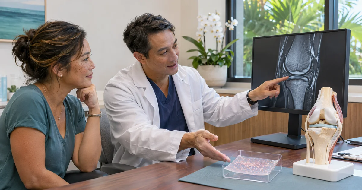
The short answer: MACI is one treatment pathway with two procedures
MACI stands for matrix-induced autologous chondrocyte implantation. It is FDA-indicated for adults with one or more symptomatic, full-thickness cartilage defects of the knee, with or without involvement of the bone beneath the cartilage. “Autologous” means the cells come from the same patient who will receive them.
The two procedures have different jobs. The first is usually an arthroscopy to inspect the knee, confirm the diagnosis, and take a small sample of healthy cartilage. The second places the cell-seeded membrane into the prepared defect. They are connected steps, but the biopsy does not obligate the patient or surgeon to proceed to implantation.
Stage one confirms more than the MRI can show
MRI helps estimate the size, depth, location, and bone involvement of a cartilage defect. Arthroscopy provides a direct view of the joint surface. The surgeon can assess whether the lesion is truly full thickness, whether its edges are contained, how much healthy cartilage surrounds it, and whether another problem—such as a meniscus tear, ligament instability, or more widespread arthritis—changes the plan.
If MACI remains a reasonable option, a small cartilage biopsy is collected from a lower-load area of the knee. The biopsy is sent for manufacturing. Other findings may be treated during that arthroscopy when appropriate, or may require a separate plan. The operation should answer a decision question, not merely harvest cells.
What happens to the cartilage sample
The biopsy contains chondrocytes, the cells that make cartilage matrix. During manufacturing, the cells are isolated and expanded, then seeded onto a resorbable porcine-collagen membrane. The final membrane is made for that patient only and is trimmed by the surgeon at implantation to match the prepared defect.
This is not a donor cartilage transplant and not a stem-cell injection. It is a regulated, patient-specific cellular product. The product must meet manufacturing and quality requirements before it can be released for use.
The interval between procedures is variable
AAOS describes MACI as a two-step process completed over several weeks, but the FDA prescribing information is more precise: the time between biopsy and implantation can vary with the quality and number of cells obtained and with clinical and scheduling factors. Cells may be held in storage until the patient and surgeon agree on an appropriate surgery date.
A single calendar estimate should therefore not be treated as a promise. Insurance authorization, medical optimization, work and caregiver planning, treatment of another knee problem, and the condition of the joint can all affect timing. Ask what must be complete before an implantation date is confirmed.
A successful biopsy does not guarantee a MACI implant
The FDA-required patient counseling language states that some patients cannot proceed even after a biopsy. The sample may be insufficient, the cells may not expand successfully, or the expanded cells may not meet quality requirements. Clinical circumstances can change as well.
The knee still has to be suitable for implantation. Severe osteoarthritis, active infection or inflammation, untreated malalignment, ligament instability, meniscal deficiency, medical risk, or inability to follow the rehabilitation program may make MACI inappropriate or require another problem to be corrected first. A pause or change in plan is not a failed promise; it is part of protecting the patient from an operation that no longer fits.
What the surgeon rechecks before implantation
Before stage two, the team should confirm that the symptoms, imaging, arthroscopic findings, and treatment goals still line up. The implant site and the mechanical environment around it matter as much as the cells.
- The defect is symptomatic, full thickness, and suitable in size, location, containment, and bone condition
- The arthritis pattern is not severe or diffuse enough to make focal restoration the wrong operation
- Alignment is acceptable or will be corrected as part of the plan
- The meniscus is functional or its deficiency is addressed
- Ligament stability is adequate or will be restored
- No active infection or uncontrolled inflammation is present
- The patient can complete the operation-specific rehabilitation and temporary activity restrictions
- The manufactured product has met release requirements and the surgical date fits its handling window
What happens during implantation
The surgeon exposes and prepares the cartilage defect, removes unstable damaged tissue while preserving healthy borders, and creates a stable bed. The membrane is shaped to the defect and placed cell-side down, commonly secured with fibrin sealant according to the product instructions and surgical plan.
The FDA labeling describes implantation through an arthrotomy. Arthroscopic implantation techniques also exist and are discussed in current AAOS surgical education; whether a defect can be treated arthroscopically depends on its location, access, shape, associated procedures, and surgeon expertise. The incision approach does not change the central requirement: accurate preparation, complete coverage, stable fixation, and protection during healing.
The rehabilitation clock starts after implantation, not biopsy
The biopsy arthroscopy and the implantation have different recoveries. The prolonged graft-protection timeline begins after the cell membrane is implanted. Location matters: a femoral-condyle graft, a patellar or trochlear graft, multiple defects, and combined procedures can require different weight-bearing, brace, motion, and exercise limits.
Early rehabilitation commonly protects the graft while restoring controlled motion and quadriceps function. Progression is based on the written protocol, swelling, gait, strength, motion, and surgeon review—not the calendar alone. Return to running, pivoting, deep loaded flexion, and sport is measured in months and requires clearance. Follow the protocol for the actual defect and any combined meniscus, ligament, or alignment procedure.
Risks and limits deserve the same attention as the technology
MACI can fail to relieve symptoms, and another procedure may be needed. Reported problems include joint pain, swelling or effusion, graft overgrowth or under-fill, graft damage or delamination, tendon irritation, cartilage or meniscus injury, and osteoarthritis progression. General knee-surgery risks include infection, stiffness, wound problems, blood clots, anesthesia complications, and injury to nearby structures.
Using a patient’s own cells does not eliminate surgical or manufacturing risk, and no cartilage procedure can guarantee a return to a particular sport or prevent future arthritis. The decision should compare MACI with nonsurgical care, microfracture or other marrow-stimulation methods, osteochondral grafting, another restoration option, or arthritis-focused treatment when the joint is too diffusely worn.
Questions to ask before the biopsy
A useful consultation makes the whole two-stage pathway visible before stage one.
- What makes my cartilage defect a possible MACI indication rather than an incidental MRI finding?
- What will the first arthroscopy confirm, and what findings could change the plan?
- Where will the biopsy be taken, and what else might be treated at stage one?
- What are the realistic alternatives if the cells cannot be manufactured or I decide not to proceed?
- How will alignment, meniscus function, ligament stability, and arthritis be evaluated?
- What must happen between biopsy and implantation, and which timing factors are outside anyone’s control?
- Would implantation be open, arthroscopic, or determined by the defect at surgery?
- Which rehabilitation protocol would apply to my defect location and any combined procedures?
- What costs, authorization steps, time away from work, transportation, and home support should I plan for?
- What outcomes are realistic for pain, function, sport, and the possibility of later surgery?
Call promptly for concerning symptoms
After either procedure, contact the surgical team for fever with worsening knee redness or heat, increasing drainage, wound opening, uncontrolled swelling, new numbness or weakness, a fall, a locked knee, or pain that suddenly escalates. New calf swelling or pain also needs prompt assessment.
Call 911 for chest pain, severe shortness of breath, fainting, or stroke symptoms. These warnings are not unique to MACI, but staged treatment means patients should know which team to contact after each procedure.
The bottom line
MACI is not simply a cartilage biopsy followed automatically by implantation. It is a staged decision pathway: confirm the defect, obtain and expand the patient’s cells, recheck the whole knee and the patient’s readiness, then implant only when the product and the clinical plan both remain suitable.
Review the complete cartilage restoration guide, compare the location-specific MACI rehabilitation protocols, or schedule an orthopedic consultation for an individualized review. Recommendations, timelines, eligibility, and outcomes vary by patient.
Sources and further reading
Medically reviewed by Paul Norio Morton, MD, FAAOS, FAAHKS. Last reviewed: . This article is for education only and isn’t a substitute for an exam and personalized advice — schedule a consultation to discuss your situation.